If you’re an ultrasound geek these tips are da bomb
SCAT 5 and concussion statement
Forwarded on behalf of Dr Rich Trenholm to us….
http://bjsm.bmj.com/content/bjsports/early/2017/04/28/bjsports-2017-097506SCAT5.full.pdf
Process Improvements in The Emergency Department
A good read from an Ontario ED
Decision fatigue
Hi all,
Really useful summary of points about MD decision fatigue in EM rap this month….
In case you don’t subscribe I’ll post in both ER’s.
I think alot of practice tips that many of us live by and may or may not voice to each other.
The days I try to live by more of these the more I enjoy what I do and feel that I am actually providing good care.
Group hug now.
John
EDTU X Resuscitation
Hi all,
Mark Mensour brought this info forward for all…
take a look!
Caep new course!
Bedside US For The Win?
We’ve been talking a lot about things we can do to improve efficiency/flow in our emergency department and I was thinking that there is a role for bedside sonography to help. I thought maybe this could stimulate discussion. I don’t expect everyone to agree with me. In general bedside sonography costs the MD some time while usually saving patients time in the department, which can still potentially help flow. In no particular order:
- In a patient with a history of prior ureteral colic, there are a few goals of care. First, analgesia. Then rule out a complicated presentation (ie septic patient), then rule out severe hydronephrosis. You don’t need a CT to rule out hydronephrosis and you don’t even need a formal scan since we know appropriately trained ED docs can diagnose hydronephrosis. We know from 1st trimester and focused RUQ bedside US studies that doing the scan yourself saves patients on average 180 minutes in the department. So it may cost you time but should help flow. And if you have a pain free patient without severe hydronephrosis they can go home. And even if you think they still need formal sonography (I’d argue they don’t) it could be done as an outpatient.
- Patients with RUQ pain. This is a no-brainer for me. Bedside US saves patients 180 minutes in the ED on average. It is EASY to look for stones and again we know ER studies of US show we are really good at it if trained appropriately. The diagnosis of cholecystitis is a CLINICAL one, not an ultrasound one, so continue to be good clinicians and you won’t miss this diagnosis. If you get a patient comfortable re-pain and prove they do or do not have a stone, they can go home. Again, if you feel they need a formal scan (I think they do not) this can be done as an outpatient with PCP follow up.
- AAA. You do not need a formal US or a CT to rule out AAA. Bedside US has superb +LR and -LR. Rupture cannot be diagnosed on US. Unstable patient with AAA = rupture and stable patient needs CT to rule out rupture. Save time and do your own bedside US to rule out this diagnosis.
- Pneumothorax and Hemothorax. There is excellent literature showing that bedside US has great +LR and -LR for these diagnoses. In fact for a supine (ie trauma) patient US preforms much better than plain films and very similarly to CT for these diagnoses. I think it probably could be argued that when the department is not busy maybe an Xray can happen as fast but I’d guess most times this is not the case.
- FAST exam in trauma. In unstable patients there is evidence that patients get to the OR quicker.
- DVT and PE. So, here is where people start getting uncomfortable. There is decent evidence that bedside compression sonography performs well compared to formal sonography re DVT and can be done in less than 3 minutes. But I have heard many MDs feel uncomfortable relying on their bedside answer for such an important diagnosis and I can’t quibble with this. You can still get a formal scan if you must but perhaps can avoid a dose of anticoagulant when we don’t have US available and while awaiting a formal scan if your compression scan is negative for DVT. Also, if you are worried about PE and bedside ECHO shows a big, hypokinetic RV then the +LR for PE is VERY high.
- Fluid overload. A big IVC, a hypokinetic LV and multiple B lines (3 or more) in multiple chest quadrants suggests LV dysfunction. The DDx for multiple B lines is anything that increases intertsitial fluid (interstitial diseases, some pneumonias, CHF, ARDS).
- Fracture reduction, especially long bones like wrist. You;ve reduced a fracture. Splint applied. Send for xray and patient comes back with an unreduced fracture. Consider doing post reduction US before applying splint. It is true they could lose the reduction in xray department but if bedside US shows the fracture is not reduced, perhaps you save some time.
There’s more but that’s all I’ve got for now. I wonder what everyone thinks? It would be nice if we had a machine that was easy to move, started up quickly, didn’t lose its battery charge immediately……
The Nitty Gritty On Stroke Thrombolysis
Sorry, but you do not have permission to view this content.
ACLS feb
Hi all,
ACLS local in feb…
Sara Tumbar in Huntsville ER asked me to post this…
Spread to anyone you want.
John
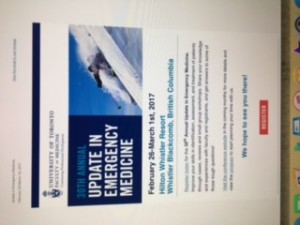
ED education in Whistler
Stroke rounds
PSR Schedule Feb 2016 -May 2018 Approved Feb 2016
Hi all,
Provincial stroke rounds may be of interest to some…see the attached schedule and host site…!
cheers
John
